Introduction
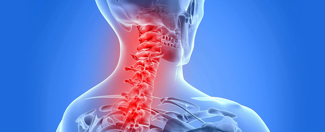
The cervical spine constitute the spinal column in the neck region which consists of seven vertebrae and their intervertebral discs in between. The intervertebral discs do not exist between the the first two cervical vertebra (atlas and axis). As most bending motion in the cervical spine occurs at the C4-5, C5-6 and C6-7 levels, disc herniation, cervical canal stenosis and bone spurs occur typically at those lower levels.
Cervical disc herniation includes soft disc herniation and/or hard disc protrusion, the latter is often associated with bone spurs build-up along the spine edges and occurs in older ages. The bone spur or disc may compress the central cervical canal directly putting pressure on the spinal cord itself and nerve roots that supply the arms at the corresponding level. This typically results in neck pain, arm pain or weakness.
Central pressure on the spinal cord may result in symptoms such as spasticity, leg weakness or gait disturbances and even walking difficulty. Surgery is advised when patients show no improvement within an appropriate time of conservative treatment.
Endoscopic surgery of the neck
Traditionally open surgery has been the main treatment for cervical spine pathology. Unlike endoscopic procedures, open surgeries with bone fusion or metal plate-screw/cage implantations partly eliminate functioning motion in the neck and cause a disturbance in the biomechanics of the cervical spine motion segment, associated with an inherent potential for long-term pain and discomfort.
Minimally invasive endoscopic cervical spine surgery is performed via a small skin incision at the front or less commonly the back of the neck. The endoscope is introduced at the targeted level and a small foraminotomy keyhole is then made at the side of the affected cervical level and the herniated disc material and any compressing bone spurs are removed using specialized miniature instrumentation, thus relieving pressure from over the spinal cord or affected nerve root. Following surgery, patients can start moving their neck normally and can typically return home on the second day.
If severe cervical spondylosis is present and most of the compression on the spinal cord is from the back or there is difficulty in accessing the spine from the front for any reason, posterior endoscopic cervical laminotomy could be performed rather than a posterior traditional laminectomy to provide extra-room for the cervical cord/nerves posteriorly or to resect a herniated cervical disc.
Benefits
Endoscopic cervical surgery is disc preserving, anatomy-maintaining and function preserving. The majority of disc material and bone is left undisturbed preserving the normal spine motion. Recovery is quick, with most patients being able to do routine daily activities starting the second day of surgery. Postoperatively, patients do not wear a cervical collar and will have normal neck motion immediately.
This endoscopic technique accomplishes efficient enlargement around the spinal cord or its nerves and maintains the structural strength of the cervical spinal column while avoiding bone fusion or metal plate-screw implantation as to maintain neck motion. Patients can usually be discharged home the following day without the need for a neck collar.
Endoscopic Operation
Animation


